

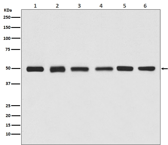
Western blot analysis of beta Tubulin expression in (1) Jurkat cell lysate, (2) Human kidney lysate, (3) 3T3 cell lysate, (4) Mouse brain lysate, (5) C6 cell lysate, (6) Rat heart lysate (H01857). Electrophoresis was performed on a 5-20% SDS-PAGE gel at 70V (Stacking gel) / 90V (Resolving gel) for 2-3 hours. The sample well of each lane was loaded with 50ug of sample under reducing conditions. After Electrophoresis, proteins were transferred to a Nitrocellulose membrane at 150mA for 50-90 minutes. Blocked the membrane with 5% Non-fat Milk/ TBS for 1.5 hour at RT. The membrane was incubated with rabbit anti-TUBB3 monoclonal antibody (Catalog # H01857) overnight at 4°C, then washed with TBS-0.1%Tween 3 times with 5 minutes each and probed with a goat anti-rabbit IgG-HRP secondary antibody at a dilution of 1:10000 for 1.5 hour at RT. The signal is developed using an Enhanced Chemiluminescent detection (ECL) kit (Catalog # EK1002) with Tanon 5200 system. A specific band was detected for TUBB3
Western blot analysis of beta Tubulin expression in (1) Jurkat cell lysate, (2) Human kidney lysate, (3) 3T3 cell lysate, (4) Mouse brain lysate, (5) C6 cell lysate, (6) Rat heart lysate (H01857). Electrophoresis was performed on a 5-20% SDS-PAGE gel at 70V (Stacking gel) / 90V (Resolving gel) for 2-3 hours. The sample well of each lane was loaded with 50ug of sample under reducing conditions. After Electrophoresis, proteins were transferred to a Nitrocellulose membrane at 150mA for 50-90 minutes. Blocked the membrane with 5% Non-fat Milk/ TBS for 1.5 hour at RT. The membrane was incubated with rabbit anti-TUBB3 monoclonal antibody (Catalog # H01857) overnight at 4°C, then washed with TBS-0.1%Tween 3 times with 5 minutes each and probed with a goat anti-rabbit IgG-HRP secondary antibody at a dilution of 1:10000 for 1.5 hour at RT. The signal is developed using an Enhanced Chemiluminescent detection (ECL) kit (Catalog # EK1002) with Tanon 5200 system. A specific band was detected for TUBB3
Anti-beta Tubulin Rabbit Monoclonal Antibody, HRP Conjugated

H01857
Product group Antibodies
Overview
- SupplierBoster Bio
- Product NameAnti-beta Tubulin Rabbit Monoclonal Antibody, HRP Conjugated
- Delivery Days Customer9
- CertificationResearch Use Only
- UNSPSC12352203