

Proteins / Signaling Molecules
This product group contains both native proteins and recombinant proteins expressed and purified from bacterial, insect and mammalian expression systems. Bio-Connect also offers fusion proteins, peptides, enzymes, bacterial and viral proteins, hormones and growth factors. Furthermore, it includes chemokines and cytokines, collagenases, albumins, globulins, blood/plasma related proteins and more.
Within the proteins/signaling molecules product group you will find many categories to find the product you need quickly. Click on one of the categories for an overview of our products.
If you need a specific product and can’t find it in our webshop, please contact our technical support.

Product group Proteins / Signaling Molecules
- SizePrice

Product group Proteins / Signaling Molecules
- SizePrice

Product group Proteins / Signaling Molecules
- SizePrice
Product group Proteins / Signaling Molecules
Protein IDO14983
- SizePrice
Product group Proteins / Signaling Molecules
Protein IDP16615
- SizePrice
Product group Proteins / Signaling Molecules
Protein IDP35354
- SizePrice
Product group Proteins / Signaling Molecules
Protein IDQ14790
- SizePrice
Product group Proteins / Signaling Molecules
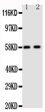
Anti-12 Lipoxygenase AntibodyPA1485-1
Protein IDP0CG47
- SizePrice
Product group Proteins / Signaling Molecules
Protein IDP04745
- SizePrice
Didn't find what you were looking for?
Search through our product groups to find the right product
Back to overview